2.3.3 Columns/Ovens
Separation columns are the heart of the GC and are housed in a temperature controlled and temperature programmable oven that can control temperatures to within 0.5 C. Considerable advances were made in gas chromatography in the 1980s, especially with regard to columns. Prior to the advent of capillary columns, chromatographic systems used packed columns. Packed columns are 1/8 to 1/4 inch metal, Teflon, or glass tubes filled with an inert resin coated with the stationary phase. Early stationary phases were highly viscous, non-volatile liquids that interact with the analytes to achieve separation or were molecular sieves that separated the analytes by molecular size and diffusion. In the early 1980s, packed columns were mostly replaced with capillary columns that are open tubular columns (internal diameters in the tenths of millimeters) with the stationary phase placed on the column walls. Initially, stationary phases were simply applied to the walls as non-volatile liquids, however today most phases are covalently bonded to the fused silica wall which yields more thermal stability. Capillary columns have dramatically more theoretical plates than packed columns and greatly improve resolution. The reader should recall this relationship from Example 1.1 in section 1.2. For example, capillary columns can have as many as 1000 times more theoretical plates as compared to a packed column. Figure 2.11 shows several GC columns.
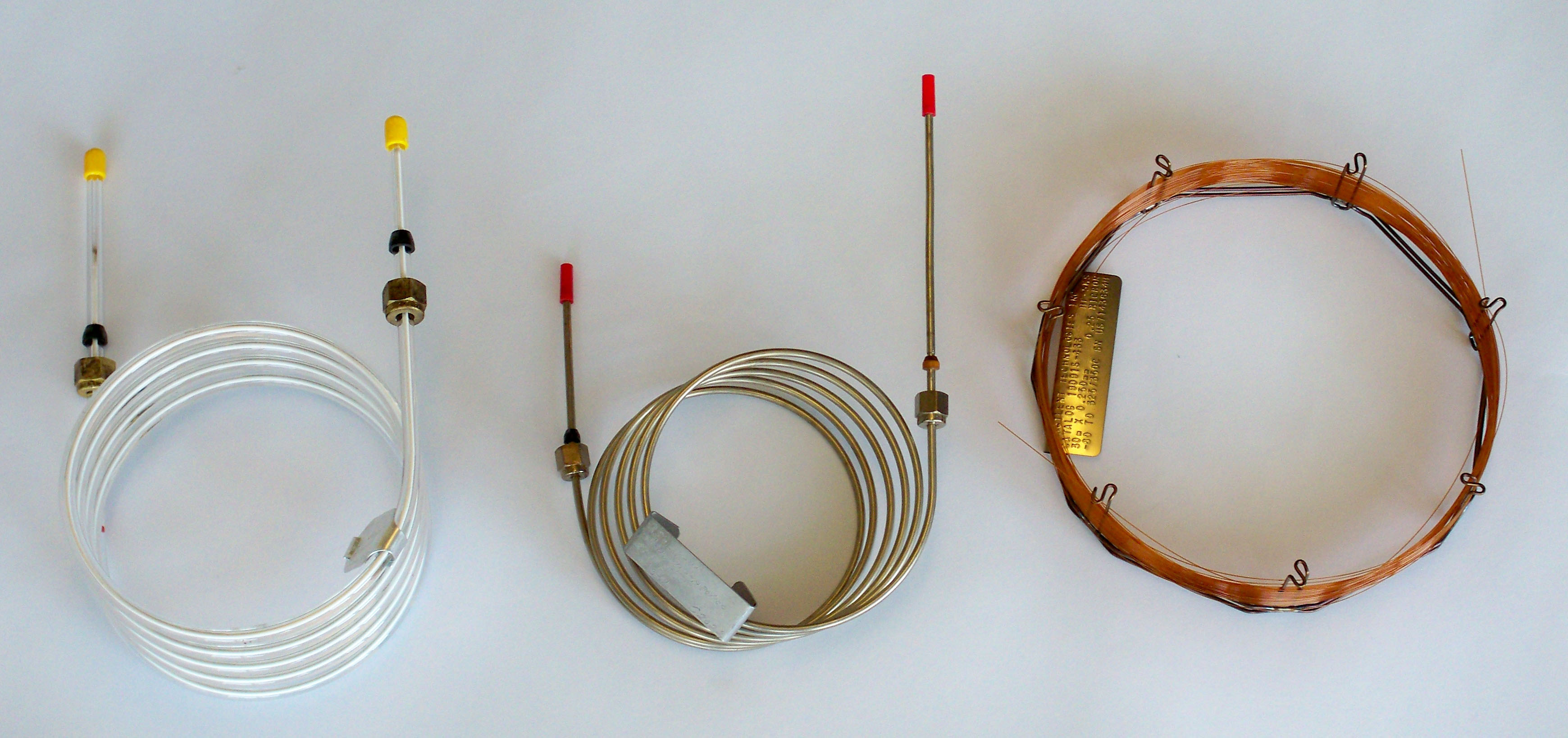
Figure 2.11 A 1/4-inch Glass Packed GC Column, a 1/8-inch Stainless Steel GC, and a Fused Silica Capillary Column (from left to right).
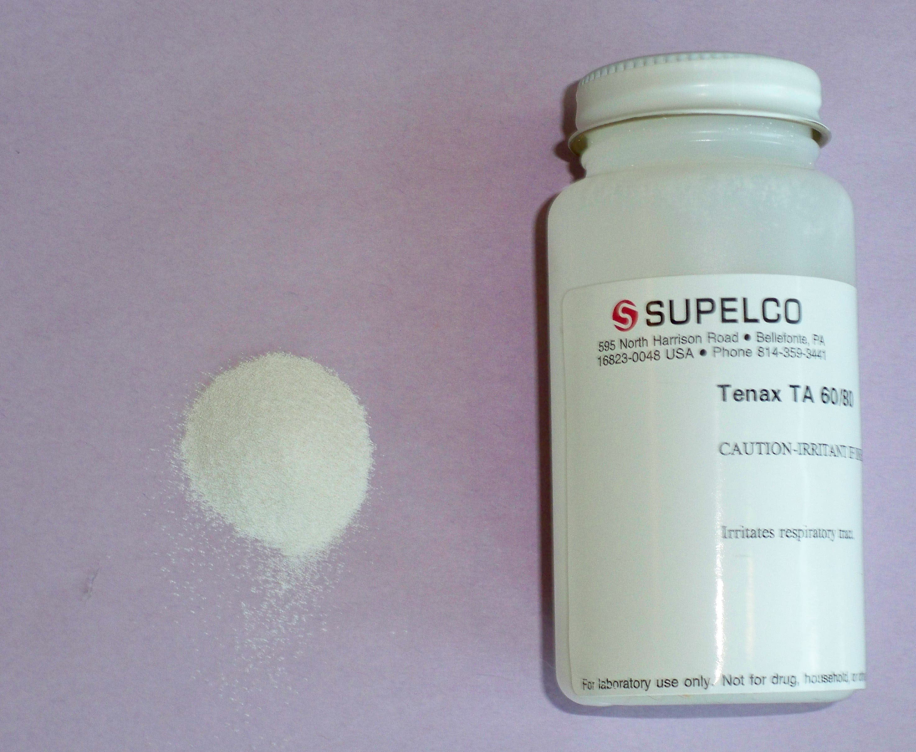
Figure 2.12 Resin for GC Packed Column.
A summary of the most common stationary phases is given in Table 1.1. The selection of a particular phase depends on the intermolecular interactions expected for the analyte of interest. As discussed in Chapter 1, the selection of the phase follows the adage “like dissolves like” or in this case “like stationary phases attach to like chemicals.” The uses of each resin are also shown in Table 1.1. Additional stationary phases are available for a variety of analyte applications. Even some chiral compounds can be separated with specialty stationary phases.
Table 1.1. Common Stationary Phases and Their Primary Use
| STATIONARY PHASE | APPLICATIONS |
|---|---|
| Polydimethyl siloxane (Trade names are DB-1, HP-1, OV-1, and SE-30) |
This is a general purpose nonpolar phase for separating hydrocarbons, polynuclear aromatics, nonpolar drugs, chlorinated pesticides, and PCBs |
| Poly(phenylmethyldimethyl) siloxane (5-10% phenyl) (Trade names are DB-5, HP-5) |
Still mostly nonpolar but with some polarity. Used to separate fatty acid methyl esters, alkaloids, drugs, and halogenated chemicals |
| Poly(phenylmethyl) siloxane (50% phenyl) (Trade names are DB-15 and OV-17) |
Slightly more polar. Used to separate more polar drugs, pesticides, and glycols |
| Poly(tritluoropropyldimethyl) siloxane (Trade names are DB-210 and OV-210 More polar. | Used to separate chlorinated aromatics nitroaromatics, and alkyl-substituted benzenes |
| Polyethylene glycol (Trade names are DB-WAX and Carbowax) |
The glycol functional group makes this phase considerably polar. Used to separate free acid, alcohols, ethers, essential oils, and glycols |
| Poly(dicyanoallyldimethyl) siloxane (Trade names are DB-1701 and OV-275) |
The most polar phase shown here. Used to separate polyunsaturated fatty acids, free acids, and alcohols |
Today most columns are fused silica capillary columns with typical internal diameters ranging from 0.25 to 0.53 mm. Column lengths range from 5 to 100 meters. As noted in Chapter 1, the longer the column, the more theoretical plates it will contain and therefore long columns are capable of separating almost any mixture of compounds. Film thicknesses range from 0.25 to 3.00 μm with thicker films usually providing more resolution (and longer analysis times). Cross-linking of films is also common and provides more thermal stability and less “column bleed” of the stationary phase. Lower column bleed provides for a more stable detector baseline and these columns are preferred in mass spectrometer applications.
| Frank's Homepage |
©Dunnivant & Ginsbach, 2008