5.7 Three-Dimensional Aspects of GC-MS
Typical chromatographic peaks were illustrated in earlier chapters. But as each chromatographic peak enters the MS it is fragmented and separated into a series of ion fragments. When graphed together on an x, y, and z plot, the x-axis represents time and traces the arrival of each compound at the chromatographic detector and the z-axis represents the total detector response that is related to analyte concentration. The mass-to-charge spectrum of each chromatographic peak is represented by a series of lines that are parallel to the y-axis and show the arrival of molecular fragments at the MS detector. Again, detector response and concentration are represented by the height of each peak. This is illustrated for one chromatographic peak in Figure 5.22.
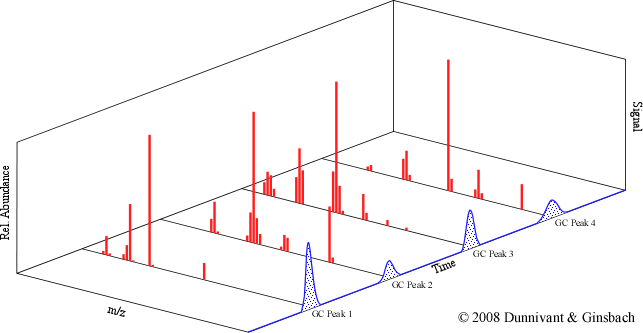
Figure 5.23. The Three-Dimensional Nature of a GC-MS Analysis.
| Frank's Homepage |
©Dunnivant & Ginsbach, 2008